
July 29, 2026
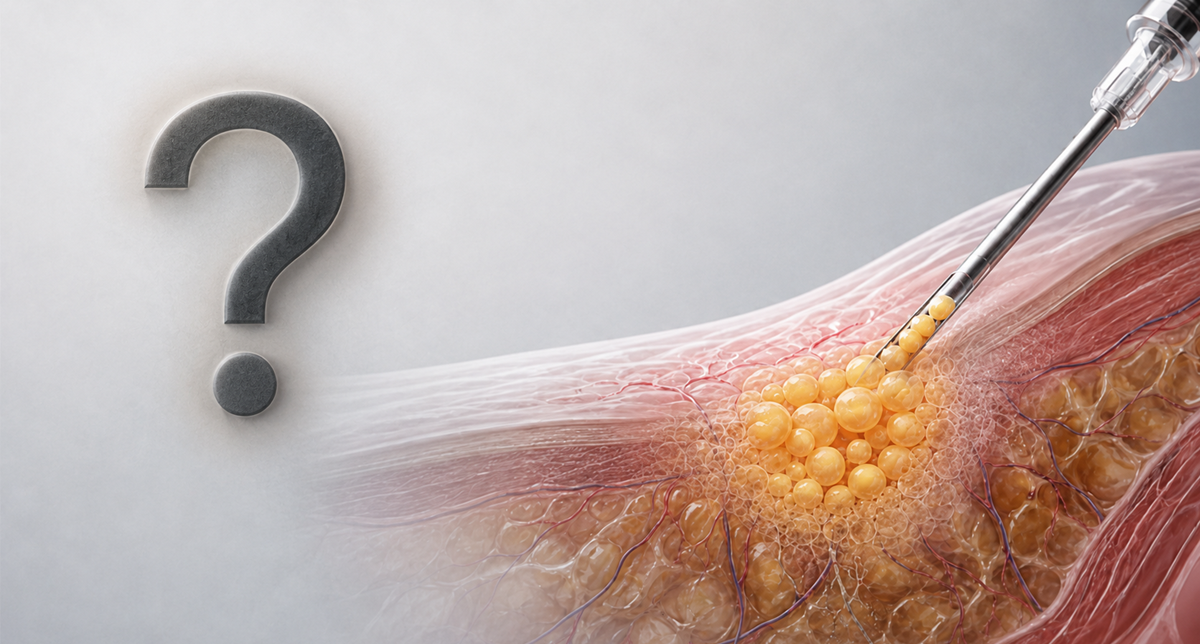 AlloClae™™ is marketed as a source of ready-to-use, donor-derived volume. It is not a conventional filler or living fat transplant, and no one has directly studied whether GLP-1 therapy changes what happens after injection.
In Part 1 of this series, we ended with an obvious question: if someone continues taking a GLP-1 medication after losing facial, breast, or buttock volume, will alloClae™-injected tissue in those areas actually persist?
It is a compelling question. It is also more complicated than it first appears, because the popular description of alloClae™ as “cadaver fat” creates the wrong biological picture.
A syringe of alloClae™ does not contain living donor adipocytes that are expected to survive as a conventional fat transplant. According to the manufacturer, the product contains no viable cells and does not depend on the metabolic activity of living cells for its primary function [1].
But calling it merely an empty scaffold is not quite right either.
The published characterization describes alloClae™ as processed human adipose tissue in which DNA and free oil are reduced while much of the tissue’s three-dimensional architecture and extracellular matrix are preserved [2]. The resulting material contains nonviable adipose structures and matrix components intended to provide immediate cushioning and support. In mice, it also showed host-cell infiltration, new blood-vessel formation and the appearance of new adipocytes over time [2].

The word retention sounds straightforward, but it can describe several biologically different outcomes:
These outcomes are not interchangeable, and a photograph showing that an area still looks fuller cannot tell us which one occurred.
In the alloClae™ characterization study, implants remained detectable over six months in athymic mice. The researchers observed host-cell infiltration, blood-vessel formation and newly forming adipose tissue, particularly by three months [2]. But this model has notable translational limits. Athymic mice retain B cells, but are T-cell deficient, so immune tolerance is only partially modeled. Implants were also placed via surgical incision rather than the cannula-based injection used clinically, so persistence in this model may not generalize to how the material behaves once injected into fibrous, vascularized human tissue. So, while the research findings support the biological plausibility of tissue integration [2], they cannot tell us how much volume will remain in a human breast, buttock or hip over several years.
They also do not tell us what any retained volume consists of: the original processed tissue, newly generated host adipose tissue, extracellular matrix, fibrosis or some combination of these components? Answering that question would require longitudinal human imaging, standardized volume measurements and, where clinically appropriate, histological analysis.
The wider literature on allograft adipose matrices offers some human experience, but even there, the research is limited. A 2026 systematic review found only 10 human studies involving 93 patients across a wide range of products, anatomical sites, injected volumes and indications. Reported retention ranged from 21.5% to 100%, reflecting substantial differences in the products, measurement methods and follow-up periods [3].
One frequently cited study of another injectable allograft adipose matrix reported approximately 47% retention at 16 weeks after small-volume injections into the dorsal wrist [4]. The same research reported 44 ± 16% retention at 24 weeks in a mouse model [4]. These are legitimate findings, but they involve a different matrix and shouldn’t be read as evidence for a substantially different product.
A small subcutaneous injection in the hand or wrist also isn’t biologically or mechanically equivalent to dozens of milliliters distributed through breast or buttock tissue, where vascular supply, mechanical loading, and tissue composition differ significantly.
The existing allograft literature therefore establishes a proof of concept: processed adipose matrices can support host-cell infiltration and adipose remodelling. It does not establish a predictable retention rate for alloClae™ across patients, doses and anatomical sites.

Volume-retention data mostly come from autologous fat grafting, where harvested fat cells (adipocytes) initially survive by getting oxygen and nutrients from nearby tissue until new blood vessels grow (vascularization). The cells farthest from that supply are most likely to die (ischemic death). Studies describe overlapping zones of survival, regeneration, and necrosis, with inadequate early vascularization driving volume loss, oil cysts, calcification, and fat necrosis. This explains why recipient vascularity, injection volume, droplet size, and technique matter so much in fat transfer [5,6].
AlloClae™, however, is biologically different: it contains no viable donor adipocytes that must survive an early ischemic window [1]. So the autologous graft literature cannot tell us if alloClae™ faces the same early cellular deadline or fails the same way. What it does suggest is that the environment still matters since vascular ingrowth supplies the oxygen, nutrients, immune cells and progenitor cells needed for remodelling. The alloClae™ mouse study showed new vessel formation and host-cell infiltration over time, but not how fast this occurs, how it scales with volume, or how it affects long-term human retention [2].
For living fat grafts to stay, the question is about adipocyte survival. For alloClae™, it’s whether the body integrates, remodels and replaces, or ultimately fails to replace the implanted tissue structure. That process has not yet been mapped in humans.
In addition to producing substantial weight loss, GLP-1 receptor agonists also exert direct and indirect effects on adipose tissue biology. It therefore seems reasonable to ask whether the drug could oppose the processes that may contribute to long-term remodelling of graft matrices.
To date, this question remains untested in humans, not just with alloClae™, but with any injectable adipose matrix, including autologous fat grafts [7].

Some evidence suggests semaglutide may inhibit adipose-tissue formation: in obese mice it altered PPAR-related lipid-metabolism proteins [8], in diabetic mice it reduced key adipogenic genes while activating AMPK and suppressing Akt [9], and in in vitro experiments with human adipocytes, it altered fat-cell-generation genes [10]. These findings hint that incretin therapy could affect graft-site fat formation, but none show this happening around an alloClae™ implant in a person.
The limitations are significant: the animal data come from visceral fat in obese/diabetic mice, not human subcutaneous implant sites, with different doses and metabolic states. Importantly, transcription-factor changes don’t necessarily predict clinical volume loss, and implant visibility may reflect persistent structural material rather than new fat formation.
Framing all GLP-1 patients as uniformly in “negative energy balance” is also an oversimplification. A patient may be actively losing weight, weight-stable on maintenance treatment, reducing the dose or regaining weight. Each distinct metabolic state, and active weight loss itself, may matter more than drug presence alone. Notably, one human study found diet-induced weight loss actually improved preadipocyte differentiation capacity, showing the effect isn’t simply “GLP-1 blocks fat growth” [11].
The biology isn’t a binary switch labelled fat burning or fat building. It is a changing system shaped by energy balance, insulin sensitivity, inflammation, adipocyte size, tissue location, dose, duration of therapy and weight loss stage.
GLP-1 therapy may not be completely hostile to graft retention: alongside any anti-adipogenic signals, it also shows favorable vascular effects relevant to graft survival.
In cultured human endothelial cells, GLP-1 promoted proliferation and tube formation supporting the idea that it may help blood vessels repair and grow [12]. Clinical and mechanistic studies have also associated GLP-1 receptor agonists with improved endothelial function and vascular health in people with metabolic disease [13].
Vascular access matters for host-cell infiltration and remodeling, but these studies don’t demonstrate improved vascularization of an adipose allograft. Endothelial tube formation in culture isn’t the same as functional vessel formation throughout an implanted tissue volume, and systemic cardiovascular benefit doesn’t guarantee local graft integration.
The published evidence points in several possible directions:
These outcomes can’t be predicted from available evidence, which is exactly why GLP-1 impacts on adipose grafts (tissue or matrix) needs to be studied in humans.

There is no evidence to date that GLP-1 therapy will cause alloClae™ to fail.
But there is also no evidence that alloClae™ behaves as a durable filler regardless of a patient’s metabolic state.
What the evidence DOES support is narrower:
What remains after alloClae™ injection, and whether ongoing GLP-1 therapy or active weight loss changes that trajectory, is unknown.
That uncertainty is not an argument against the technology. It is a research question hiding inside a rapidly growing clinical market.
In our third and final part of this series, we turn from the science of alloClae™ to an equally unsettled system: the laws governing human tissue. There, the question is no longer whether the volume stays, but how a product made from donated adipose tissue should be classified, and why a New York statute with roots in a notorious body-snatching scandal has become the obstacle in its path.
Stay tuned for Part 3: “The Law Was Written for Grave Robbers.”
This article is educational and non-promotional. It reflects publicly available reporting, manufacturer information, regulatory guidance and peer-reviewed literature as of July 2026. Clinical, regulatory and legal information concerning alloclae™ continues to evolve and should be verified against current primary sources before reliance. Individual patient accounts are illustrative and do not establish population-level incidence or causation.
References